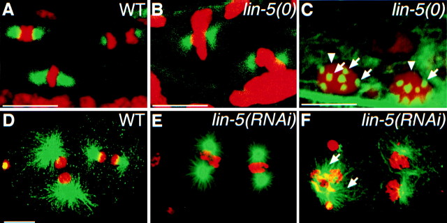
Figure 7.
Immunostaining of tubulin, showing that lin-5 is not essential for bipolar spindle formation, centrosome duplication, and microtubule nucleation, but is required for correct spindle positioning. Tubulin staining is in green, DNA staining in red. Bipolar spindles formed in intestinal nuclei of wild-type (A) and lin-5 mutant (B) L1 larvae. We also observed apparently normal bipolar spindles in lin-5 mutants in the Q, V, H, and P cells (data not shown). C, Late L1 lin-5 mutant with multiple centrosomes (arrows) and polyploid nuclei (arrowheads) in the ventral nerve cord. The number of centrosomes was found to double with each defective division. D, A wild-type embryo showing normal spindle formation and positioning at the two-cell stage. Anterior is to the left. E, A lin-5(RNAi) two-cell embryo showing apparently normal spindle formation, but defective spindle rotation. The spindle of the P1 blastomere did not rotate to establish a longitudinal spindle orientation, in contrast to that in the wild-type embryo in D. F, A lin-5(RNAi) embryo in which spindle microtubules were formed (arrows), even subsequent to defective nuclear and cytoplasmic division of the AB cell (left). Note that the first rounds of chromosome congression and segregation are completed in lin-5(RNAi) embryos, but not in lin-5(0) mutant larvae. Bars, ∼10 μm.

