
Figure 8.
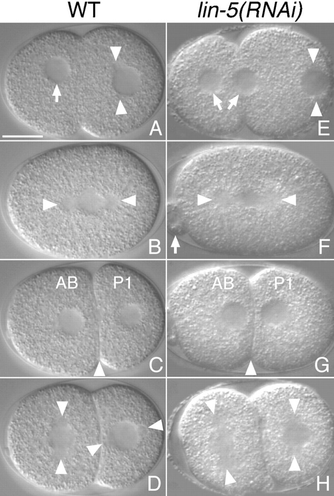
Nomarski images of living embryos. A–D, Selected images from a chronological time-lapse sequence showing early division events in a wild-type embryo. E–H, Images from multiple lin-5(RNAi) embryos illustrating defects in meiotic division and mitotic spindle positioning. Anterior is to the left, ventral is down. A and E, Pronuclear migration and pseudocleavage. During meiosis in the wild-type, a single maternal pronucleus is formed (arrow) that migrates to the posterior. Meiotic defects can result in the formation of two maternal pronuclei (arrows) in lin-5(RNAi) embryos. Arrowheads indicate the sperm pronucleus with duplicated centrosomes. B and F, Formation of the first mitotic spindle. Asters are indicated by arrowheads; arrow points to an abnormally large polar body in the lin-5(RNAi) embryo. C and G, Two-cell embryos. The lin-5(RNAi) embryo has divided symmetrically. Arrowheads indicate cleavage plane. D and H, Preceding the second divisions, the centrosome/nucleus complex has rotated in the wild-type P1 blastomere, but not in the lin-5(RNAi) embryo. Arrowheads indicate spindle positions. Bar, 10 μm.