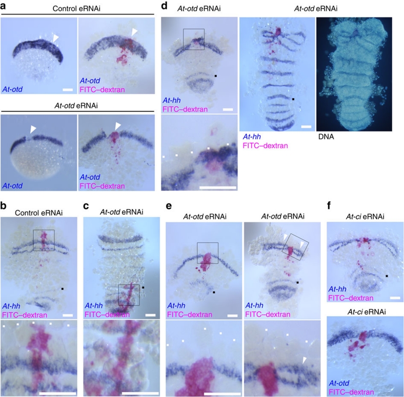
Figure 5. At-otd activity is required to maintain dynamic At-hh expression in the presumptive head ectoderm.
(a) Detection of At-otd transcripts in embryos microinjected with control gfp dsRNA (upper panels) and At-otdXE dsRNA (lower panels). Each embryo was photographed before (left) and after (right) staining for the lineage tracer FITC–dextran (pink). Arrowheads indicate areas where the lineage tracer and presumably the dsRNAs were introduced. (b) Control (gfp) eRNAi embryo stained for At-hh transcripts (purple) and the cell-lineage tracer FITC–dextran (pink), which was detected in the presumptive head ectoderm. Embryos shown in the following panels were stained in the same way as this embryo, unless otherwise indicated. (c) At-otd eRNAi embryo in which the labelled cell clone (pink) extended from the L4 segment to the caudal lobe. (d) Two At-otd eRNAi embryos that experienced having a labelled clone (pink) at the anterior margin of the nascent germ band at stages 6 and 7. The embryo in the left panel was fixed at stage 7, and that in the middle and right was allowed to develop until stage 8 and fixed for staining. The right panel shows the DNA fluorescence image. (e) At-otd eRNAi embryos in which the labelled clone (pink) was slightly removed from the anterior margin (white dots). The right embryo was slightly older than the left one. Arrowheads in the right panel indicate the adjacent At-hh stripes that each exhibited a characteristic splitting pattern with sizes adjusted to the fields. (f) At-ci eRNAi embryos; the lower one was stained for At-otd transcripts (purple) and the cell-lineage tracer FITC–dextran (pink). All embryos in this figure were at germ band-forming stages (corresponding to stage 7) except the embryo in right and middle panels of d, which was at stage 8. Boxed areas in the upper panels are magnified in the lower panels (b,c,e, left column of d). White dots indicate the anterior margin (b,d,e), and black dots indicate the At-hh stripe at the L4 segment (b–f). Scale bars, 100 μm.

