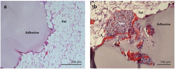
Figure 13.
Histological results from implantation of PEG-catechol hydrogel into mice. (a) Hematoxylin-eosin-stained tissue section showing the adhesive in contact with fat tissue after 6 weeks of implantation. Note the close apposition of the adhesive to the fat tissue and limited inflammatory cell infiltration. (b) Hematoxylin-eosin-stained tissue section showing a site of mouse islet transplantation using the PEG-catechol gel as a sealant to immobilize islets onto the fat tissue surface. The graft was removed on day 100 following transplantation. Revascularization of the islet is apparent from the presence of several blood vessels containing erythrocytes (red ). Adapted from Reference 73 with permission.

