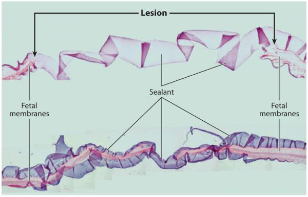
Figure 14.
Ex vivo sealing of fetal membrane defects with PEG-catechol adhesive. Through-thickness puncture wounds (sealant lines) were created on fresh fetal membranes with an ~3.5-mm trocar, and approximately 0.5 ml of adhesive was applied over the defect. The images represent a collage of a hematoxylin- and eosin-stained cross section through the defect and the PEG-catechol adhesive. The hydrogel appears as a ribbon-like structure that bridges the puncture edges. The bottom image shows a cross section of the same lesion at a narrow location. Adapted from Reference 98 with permission.

