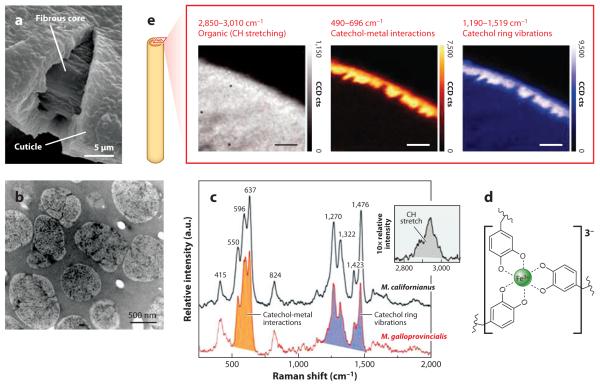
Figure 8.
The cuticle of Mytilus byssus. (a) Scanning electron micrograph of a cracked cuticle showing the thickness relative to the underlying fibers. This thread was stretched to 120% its initial length. (b) Transmission electron micrograph of a 1-μm-thick section of the cuticle in Mytilus galloprovincialis. There is a continuous homogeneous matrix filled with marbled inclusions. (c) Resonance Raman spectra taken of cuticular thin sections of Mytilus californianus (black line) and M. galloprovincialis (red line) using a 785-nm laser. (d) A tris-Fe(III)-Dopa complex. (e) Confocal Raman micrographs of byssal cuticle imaged at the C-H stretching shift (left), at the catechol-O-Fe(III) shift (center), and at the Dopa ring vibrations (right). Adapted from research originally published in Reference 38, copyright © AAAS.

