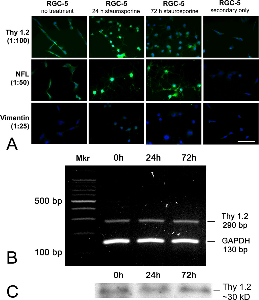
Figure 1. Detection of Thy1.2 and other neuronal markers in undifferentiated RGC-5 cells, RGC-5 cells differentiated with staurosporine for 24 and 96 h.
(A) Three retinal cell markers were examined by immunostaining: Thy 1.2, a ganglion cell marker; neurofilament-light (NF-L), a neuronal cell marker; and vimentin, a retinal glial cell marker. Bright whitish fluorescence indicates positive immunodetection of the primary antibody; light gray fluorescence indicates Hoescht 33258 staining of cell nuclei. Scale bar denotes 25 µm. (B) cDNA was isolated from RGC-5 cells cultured in the absence or presence of staurosporine for 24 or 72 h and subjected to RT-PCR using primers specific for mouse Thy 1.2 (290 bp), GAPDH was the internal control (130 bp). “Mkr” = DNA marker. (C) RGC-5 cells were grown to confluence, cultured in the absence or presence of staurosporine for 24 or 72 h, protein isolated, and subjected to immunoblotting using an antibody against Thy 1.2 (30 kD).

