Figure 1.
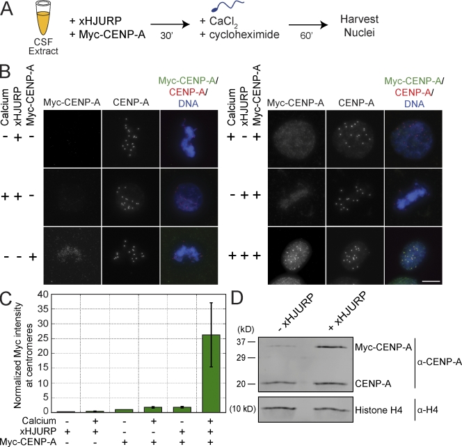
CENP-A assembly in Xenopus extracts requires xHJURP addition and mitotic exit. (A) Schematic of xHJURP-mediated CENP-A assembly assay in Xenopus egg extract. (B) Representative images from xHJURP-mediated CENP-A assembly assay. The staining for myc–CENP-A, total CENP-A, or the merge of myc–CENP-A, total CENP-A, and DNA is indicated above the images. Calcium, xHJURP, or myc–CENP-A addition is shown to the left of each image row. Bar, 10 µm. (C) Quantification of myc–CENP-A fluorescence intensity at centromeres for the assembly reactions represented in B, normalized to the metaphase control sample without xHJURP but with myc–CENP-A RNA added (−/−/+). Mean per pixel intensity at centromeres was quantified, and >200 centromeres were quantified per condition. Error bars show SEM; n = 4. (D) Western blot of chromatin fractions from CENP-A assembly reactions performed in the presence (+) of calcium, Myc-CENP-A, and xHJURP (+/+/+) or in the presence of calcium and Myc-CENP-A but lacking (−) xHJURP (+/−/+). Histone H4 was used as a loading control. n = 3.

