Figure 3.
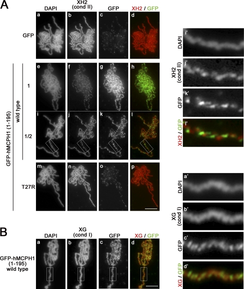
The N-terminal domain of hMCPH1 competes for chromosome binding of condensin II in Xenopus egg extracts. (A) A reticulocyte lysate containing GFP or GFP-hMCPH1 (amino acids 1–195; wild type and T27R) was mixed with 10 vol metaphase egg extracts and incubated for 30 min. Sperm chromatin was then added and incubated for another 120 min. Metaphase chromosomes assembled were fixed and stained with DAPI (a, e, i, and m), anti–XCAP-H2 (XH2; b, f, j, and n), and anti-GFP (GFP; c, g, k, and o). To reduce the dose of GFP-hMCPH1 (amino acids 1–195) to 50% (i–l), the reticulocyte lysate containing the wild-type protein was diluted twofold with a mock lysate before being mixed with the egg extracts. Close-ups of chromosomal regions indicated by the white rectangles in i–l are shown in i′–l′. (B) Metaphase chromosomes assembled in the presence of GFP-hMCPH1 (amino acids 1–195; wild type) as described in A were fixed and stained with DAPI (a), anti–XCAP-G (XG; b), and anti-GFP (GFP; c). Close-ups of chromosomal regions indicated by the white rectangles in a–d are shown in a′–d′. Bars, 5 µm. cond, condensin.

