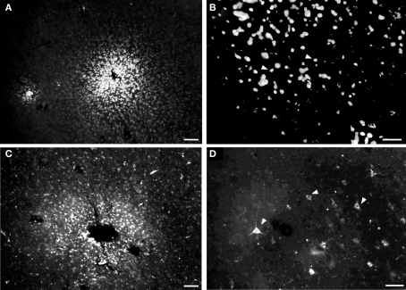
Figure 5.
Photomicrographs of injection sites of fluorescent anatomical tracers and retrogradely labeled neurons in tangential cortical sections. (A) Site labeled after insertion of a small crystal of diamidine yellow (DY). (B) Cellular nuclei retrogradely labeled by DY. (C) Fluororuby injection site. (D) Neurons retrogradely labeled by fluororuby (white arrowheads show some examples) Calibration bars: (A,C) = 50 μm; (B,D) = 100 μm.

