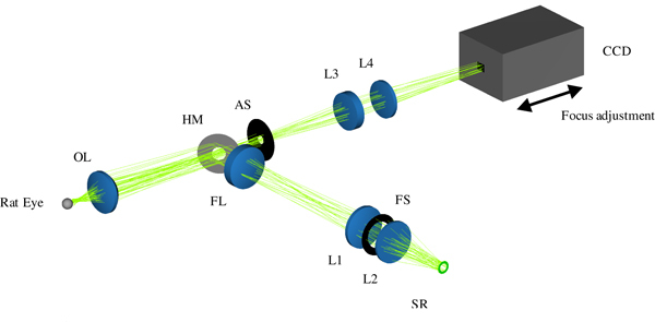
Fig. 5.

Total optical retina camera in ZEMAX NSC mode, showing the integrated lens optical setup for fundus illumination (folded path) and imaging, adapted to the rat eye. An annular light source, the reduced schematic rat eye, and a CCD are added to enable realistic ray tracing and illustration. Illumination path: source ring (SR) – lenses (L1, L2) – field lens (FL), defines the field of illumination at fundus – hole mirror (HM) – ophthalmoscopic lens (OL) – rat’s pupil (not shown). Imaging path: rat’s fundus – OL – HM – aperture stop (AS) – lenses (L3, L4) – detector. Focus adjustment is done by moving the CCD as indicated by arrows.
