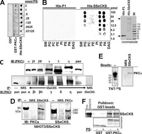
FIGURE 1.
SSeCKS association with PKC isoforms and with PS. A, SSeCKS binds PS. Shown is the overlay assay of GST, GST-PKCα, or His-TAT-SSeCKS on membranes containing dilutions of PS probed with anti-GST or anti-SSeCKS Ab. B, SSeCKS binds multiple phospholipids. Left panel, overlay assay of control His-TAT-DHRS12-P1 or His-TAT-SSeCKS protein on membranes containing dilutions of sphingomyelin (SM), phosphatidylglycerol (PG), phosphatidylcholine (PC), phosphatidylethanolamine (PE), phosphatidylinositol (PI), PS, phosphatidic acid (PA), or diacylglycerol (DAG) probed with anti-His6 mAb (right panel, Coomassie-stained gel showing the 37-kDa His-TAT-DHRS12-P1 and the 300-kDa His-TAT-SSeCKS proteins; M, marker proteins). C, SSeCKS associates with cPKC and nPKC isoforms. Upper panel, IB of SSeCKS-overexpressing NIH3T3 lysates using PKC isoform-specific mAbs; lower panel, IP of SSeCKS-overexpressing NIH3T3 lysates with anti-SSeCKS or NRS followed by IB with isoform-specific PKC Abs, pan-PKC (recognizes α, β, and γ), or SSeCKS Ab. D, reciprocal co-IP between SSeCKS and PKCα. Lysates from S24 cells (NIH3T3[Tet-Off/SSeCKS] (30)) grown in the absence of tetracycline were immunoprecipitated with NRS, anti-SSeCKS, or anti-PKCα Ab, and then the IPs were probed by IB using the reciprocal Ab. E, left panel, autoradiography of 35S-labeled full-length PKCα produced in TnT reactions; right panel, pulldown of labeled PKCα proteins using beads containing NRS or SSeCKS (after IP from S24 cell lysates). F, beads with GST or GST-PKCα (Coomassie-stained in the bottom panel) were used to precipitate His-SSeCKS protein in the presence or absence of PS (20 μg/ml) followed by SSeCKS IB (top panel).

