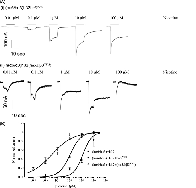
FIGURE 3.
Functional properties of (hα6/hα3)hβ2*-nAChR. A, representative traces are shown for inward currents in oocytes held at −70 mV, responding to application at the indicated concentrations of nicotine (shown with the duration of drug exposure as black bars above the traces), and expressing nAChR hα6/hα3, hβ2, and either (i) hα5V9′S or (ii) hα5/hβ3V9′S subunits. Calibration bars are 100 (i) or 50 nA (ii) currents (vertical) or 10 s (horizontal). Note the differences in inward current kinetics. B, results for these and other studies averaged across experiments were used to produce concentration-response curves (ordinate, mean normalized current ± S.E.; abscissa, ligand concentration in log μm) for responses to nicotine as indicated for oocytes expressing nAChR hα6/hα3 and hβ2 subunits alone (■), with hα5V9′S subunits (●), or with hα5/hβ3V9′S subunits (△). Current amplitudes are represented as a fraction of the peak inward current amplitude in response to the most efficacious concentration of nicotine. Leftward shifts in nicotine concentration-response curves are evident for functional nAChR containing hα5V9′S or hα5/hβ3V9′S subunits (p < 0.0001; ∼12- and 433-fold lower EC50 values, respectively). See Table 2 for parameters.

