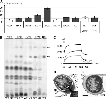
FIGURE 3.
The substrate of TolC-DevBCA. A, ATP hydrolysis rates of DevAC in presence of indicated substrate mixes. ACB represents DevAC with DevB; HCE represents DevAC, DevB, and heterocyst cell extract; HMF represents DevAC, DevB, and heterocyst membranes; HCW represents DevAC, DevB, and heterocyst cell walls; HGL represents DevAC, DevB, and purified HGLs; MCW represents DevAC, DevB, and cell walls of mutant DR181; MCM represents DevAC, DevB, and cytoplasmic membranes of mutant DR181; AC represents DevAC without DevB; MT represents DevAC and DevBN333A, +/−HGL represents with/without purified HGLs. All substrate concentrations were adjusted to 4 μg of the respective protein fraction or equal to 4 μg cell wall protein in the case of adding HGLs. Gray bars indicate the presence of HGLs in the respective fractions. ATPases possibly present inside the substrate fractions were preinactivated by incubation with 5 mm VaO4. B, thin layer chromatography of extracts of complemented mutants DR74DevB and DR74DevB_N333A, and purified HGLs. WT, DR74DevB; MT, DR74DevB_N333A; VCE, vegetative cell extract; HCE, heterocyst cell extract; HCM, heterocyst cytoplasmic membranes; HCW, heterocyst cell walls; HGL, purified HGLs. Arrows indicate HGLs. C, SPR analysis of the interaction of either immobilized TolC (TolCsol_i8H; ∼730 RU; black curves) or DevAC (DevAC_i8H; ∼510 RU; gray curves) to DevB (DevBsol; solid lines) or DevBN333A (DevBsol_N333A; dashed lines). DevB was injected in the reaction buffer at 2.0 μm. D, electron micrograph of a heterocyst of strain DR74DevB. HEP, heterocyst envelope polysaccharide layer; HGL, glycolipid layer. Error bar, 1 μm. E, electron micrograph of a heterocyst of strain DR74DevB_N333A. Bar, 1 μm.

