FIGURE 3.
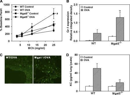
Allergen-challenged Mgat5−/− mice exhibit increased AHR and develop lung neutrophilia. A, AHR in control and allergen-challenged WT and Mgat5−/− mice assessed by whole body plethysmography. The enhanced pause in breathing after MCh was expressed as a percentage of baseline enhanced pause with saline (n = 8 mice per group). B, Gr-1 expression in lung tissue lysates of control and allergen-challenged mice by densitometry of Western blots normalized against β-actin expression (n = 4–5 per group). C, representative images of lung tissue neutrophils in allergen-challenged WT and Mgat5−/− mice by confocal microscopy after staining with mAb NIMP-R14 (magnification, ×600). No NIMP-R14-positive cells were detected in control lungs from both groups (n = 4–5 per group). D, KC levels in lung tissue lysates of control and allergen-challenged WT and Mgat5−/− mice by flow cytometry (n = 4–6 per group). Data represent mean ± S.E. in A, C, and E. *, p < 0.05 versus respective control; and #, p < 0.05 between allergen-challenged groups for A; **, p < 0.05 versus WT OVA in B and D.

