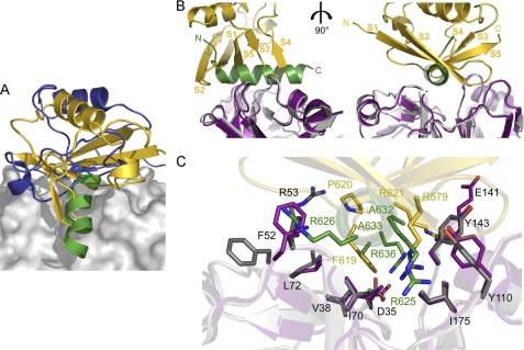
FIGURE 7.
Competitive cofactor binding to p97N. A, ribbon representations together with the molecular surface of p97N in complex with the VIM of gp78 (this study, colored in green), FAF1UBX (Protein Data Bank entry 3QQ8, colored in gold) and NPL4ULD (Protein Data Bank entry 2PJH, colored in blue). B, ribbon representations of p97 in complex with FAF1UBX (Protein Data Bank entry 3QQ8) and gp78 (this study) with p97N in magenta (FAF1UBX complex) and gray (gp78 complex), FAF1UBX in gold, and gp78 in green. Two orientations that differ by a 90° rotation are shown. C, close-up view showing the common p97N binding surface important for FAF1UBX and gp78 interaction. Key residues involved in the respective interaction are shown in a stick representation using the same color code for carbon atoms as in A.

