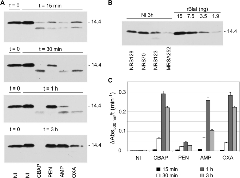
FIGURE 2.
Time course of BlaI turnover and resynthesis in whole-cell extracts, and of PC1 β-lactamase activity in the media, after induction of the bla system by β-lactam antibiotics in S. aureus NRS128 (the results for the three other strains are given in the supplemental material). A, the BlaI protein was detected in whole-cell extracts (60-μg portion of total protein) of non-induced (NI) and induced (15 min, 30 min, 1 h, and 3 h) cultures by Western blot analysis, using anti-BlaI antibody. The proteins were separated by SDS-PAGE (15% gels) and transferred to nitrocellulose membrane, and the IgG-binding proteins were blocked with human IgG and Protein A/G blocker (GenScript) prior to the immunoblot analysis. Numbers to the right indicate the position of migration of the molecular mass markers (kDa). B, comparison of the amount of BlaI in whole-cell extracts of the strains NRS128, NRS123, NRS70, and MRSA252 (60-μg portion of total protein), using purified rBlaI as a standard. The cultures were grown in the absence of antibiotics for an additional 3 h after A625 of 0.8 was reached (non-induced condition at 3 h). C, the chromogenic β-lactam antibiotic nitrocefin was used as a reporter to monitor the β-lactamase levels in the culture media of non-induced cultures and after induction with CBAP, penicillin G, ampicillin, and oxacillin for different times (15 min (black bars), 30 min (white bars), 1 h (dark gray bars), and 3 h (light gray bars). All cultures were exposed to different antibiotics at the same A value (A625 = 0.8). Error bars, S.E.

