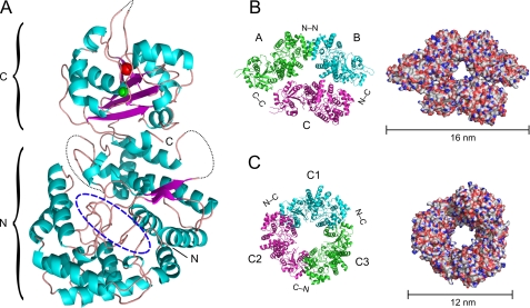
FIGURE 2.
Crystal structure of NP of LASV strain AV. A, ribbon diagram of the NP monomer. NP is composed of N- and C-terminal domains. The N-terminal domain contains a deep cavity (dashed blue oval) previously shown to bind pyrimidine nucleotides (22). The C-terminal domain contains two metal ions: Mn2+ (red sphere), coordinated by Asp-389, Glu-391, and Asp-533; and Zn2+ (green sphere), coordinated by Glu-399, Cys-506, His-509, and Cys-529. The dotted black line depicts the chain in loop regions that could not be solved. B, asymmetric trimer representing the asymmetric unit composed of chains A, B, and C (left, ribbon diagram; right, molecular surface). C, symmetric trimer composed of C chains from three asymmetric units viewed along a crystallographic 3-fold rotation axis (left, ribbon diagram; right, molecular surface).

