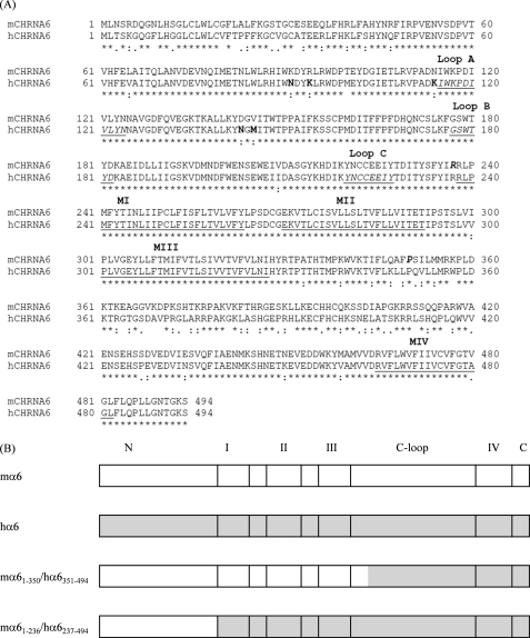
FIGURE 1.
A, sequence alignment for mouse (m) or human (h) nAChR α6 subunits (GenBankTM NP_004189.1 (Homo sapiens) and NP_067344.2 (Mus musculus); single letter code, numbering begins at translation start methionine). Symbols below sequences indicate fully (*), strongly (:) or weakly (.) conserved residues, and the lack of a symbol indicates amino acid divergence, and boldface type in hα6 subunit indicates residues given prime attention in mutagenesis studies. Underlining in the hα6 subunit sequence indicates putative transmembrane domains. Underlined and italicized type in the hα6 subunit indicates putative domains involved in ligand binding (loops A–C), and boldface and italicized type in the mα6 subunit indicates junctions for chimeric subunits. B, schematic diagrams of wild-type, human, or mouse α6 subunits or chimeric subunits. Notations are: N = N-terminal domain; I, II, III, or IV = respective transmembrane domains; C-loop = cytoplasmic loop; C = C terminus.

