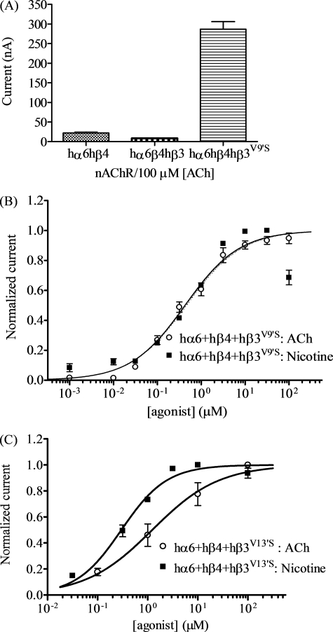
FIGURE 2.
Functional properties of hα6hβ4*-nAChR. A, mean peak inward current amplitude (±S.E.; abscissa; nA) elicited by oocytes expressing the indicated human nAChR subunit combinations in response to application of 100 μm ACh. The level of nAChR function in oocytes expressing hα6 and hβ4 subunits is reduced by addition of wild-type hβ3 subunits but increased in the presence of hβ3V9′S subunits (p < 0.05). B and C, results averaged across experiments were used to produce concentration-response curves (ordinate − mean normalized current ± S.E.; abscissa − ligand concentration in log μm) for responses to ACh (○) or nicotine (■) as indicated for oocytes expressing nAChR hα6 and hβ4 subunits with either hβ3V9′S (B) or hβ3V13′S (C) subunits. Concentration-response curves for ACh and nicotine are almost superimposable for oocytes expressing hα6, hβ4, and hβ3V9′S subunits, but EC50 values for ACh and nicotine are different (p < 0.0001) for oocytes expressing hα6, hβ4, and hβ3V13′S subunits (see Table 1 for parameters).

