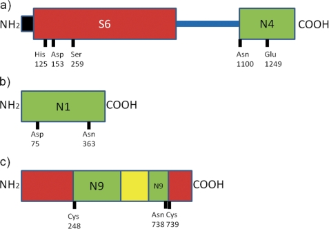
FIGURE 3.
The domain structures of (a) the Tsh precursor from Escherichia coli (family N4), (b) the coat protein from flock house virus (family N1), and (c) the intein-containing V-type proton ATPase catalytic subunit A from S. cerevisiae (family N9) are shown. Domains are shown as rectangles on a cyan string. The signal peptide is shown in black; the asparagine peptide lyase domain in green and other domains in red (the serine peptidase domain for the Tsh precursor and the extein domains for the RecA protein) and yellow (the homing endonuclease within the ATPase intein). Active site residues are indicated along the bottom edge of each domain.

