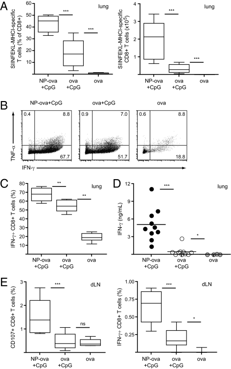
Fig. 2.
Lung effector CD8+ T-cell responses are increased following pulmonary immunization with NP-ova and CpG. Mice were immunized as described in Fig. 1. (A) Proportion (Left) and total numbers (Right) of ova-specific CD8+ T cells in the lung was determined by pentamer staining on day 19. (B and C) The proportion of IFN-γ- and TNF-α-producing CD8+ T cells was determined after 4 h restimulation of lung leukocytes with PMA/ionomycin by intracellular staining and flow cytometry. Dot plots are gated on CD8+ T cells; values represent the percentage of cells detected in each gate. (D) Lung leukocytes were isolated and cultured for 4 d in the presence of SIINFEKL; IFN-γ secretion in the supernatant was determined by ELISA. (E) Lung draining LN cells were isolated on day 19 and restimulated ex vivo for 6 h in the presence of SIINFEKL; CD107a and IFN-γ expression by CD8+ T cells was determined by flow cytometry. Data in scatter plots represent values from single animals. Experiments were repeated with 10 mice per group.

