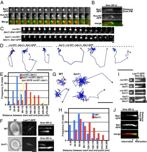
Fig. 5.
Bzz1 associates with and stabilizes the base of endocytic membrane invaginations. (A) Dynamic localization of Rvs167-RFP relative to Bzz1-GFP in living cells. The time series shows the composition of individual patches from two-color movies (1 frame per second). White dashed lines in merged images represent the plasma membrane (PM). (Scale bar: 300 nm.) (B) Kymograph representation of Rvs167-GFP and Bzz1-RFP in a single patch from a two-color movie. Kymographs are oriented with the cell exterior at the top. (Scale bar: 400 nm.) (C) Displacement of Sla1-GFP from its starting position on retraction to the PM in rvs167Δ bbc1Δ and bzz1Δ rvs167Δ bbc1Δ mutants. The time series shows the position of individual patches from single-color movies (1 frame per second). Arrowheads represent starting positions. (Scale bar: 200 nm.) (D) Tracking of individual Sla1-GFP patches in rvs167Δ bbc1Δ and bzz1Δ rvs167Δ bbc1Δ mutants. The positions of the centers of patches were identified in each frame of a movie (1 frame per second) from the medial focal plane of a cell. Consecutive positions from the start (green) to the end (red) were connected by lines. Patch traces are oriented so that the cell surface is up (dashed line) and the cell interior is down. The time difference between each position along the track is 1 s. (Scale bars: 200 nm.) (E) Histogram shows the distribution of distances between the appearance and disappearance sites for retracting Sla1 patches in the rvs167Δ bbc1Δ, bzz1Δ rvs167Δ bbc1Δ, and bzz1-R37E rvs167Δ bbc1Δ mutants. (F) TIRF microscopy analysis of Las17-GFP dynamics on the PM in living cells. The single frame was taken during bright-field (BF) and GFP fluorescence single-color imaging. A kymograph representation of Las17-GFP in live-cell movies (1 frame per second) in WT cells (Upper) and bzz1Δ (Lower) mutants is shown. White dashed lines indicate the regions used for kymographs. (Scale bar: 2 μm.) (G) Tracking of two individual Las17-GFP patches from TIRF images. Positions of the centers of patches were determined in each frame of a movie (1 frame per second). Consecutive positions from the start (green) to the end (red) are connected by lines. (Scale bar: 100 nm.) (H) Histogram shows the distribution of distances between appearance and disappearance sites for Las17-GFP imaged using TIRF microscopy. (I) Kymograph representation from TIRF imaging of Las17-GFP sliding along the PM in rvs167Δ, bzz1Δ rvs167Δ, rvs167Δ bbc1Δ, and bzz1Δ rvs167Δ bbc1Δ mutants from movies (1 frame per second). (Left) Images are the first frames of each movie. Kymographs were obtained from a single patch for each mutant. White dashed lines represent the PM. (Scale bar: 400 nm.) (J) Kymograph representations of Bzz1-GFP and Sla1-RFP of a single patch in an rvs167Δ mutant from a two-color movie of a medial focal plane in wide-field microscopy (1 frame per second). (Scale bar: 400 nm.)

