Fig. 4.
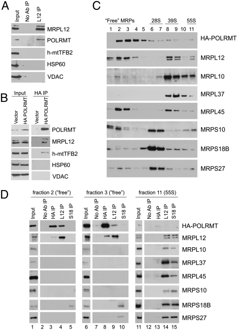
Identification of a “free” pool of MRPL12 that selectively interacts with POLRMT. (A) Immunoblotting of proteins that co-IP with MRPL12 from HeLa whole-cell extracts. IP with no antibody added (No Ab IP) was used as a negative control. (B) Western blot analysis of proteins recovered in HA antibody IP (HA IP) from HeLA cell expressing HA-tagged POLRMT (HA-POLRMT) or empty vector as a negative control (vector). (C) Sedimentation of HA-POLRMT-expressing HeLa cell extracts through 10–30% sucrose gradients followed by Western blot analysis. Fraction numbers (1-lightest, 11-heaviest) are shown. Fractions enriched for “free” MRPs, small (28S) and large (39S) ribosomal subunits, and fully assembled ribosomes (55S) are underlined. (D) Western blot analysis of proteins that co-IP with HA-POLRMT, MRPL12 and MRPS18B antibodies. Fractions 2, 3, and 11 from (C) were used for pull-down (input). IP with no antibody added (No Ab IP) was used as a negative control. In all boxes, antibodies used for Western blot analysis are indicated.

