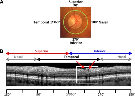
Figure 1.
(A) The locus of the fdOCT circle scan (green circle) is shown with a 360° scale to indicate disc location. 0° is the most temporal part of disc and occurs at 9 o'clock for a right eye and 3 o'clock for a left eye. (B) Circle scan of eye 33. White box: region with holes, enlarged in Figure 2A.

