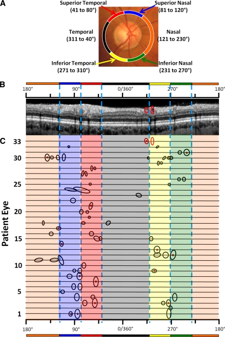
Figure 4.
Location of holes by patient. (A) Garway-Heath et al.7 disc sectors. (B) The same circle scan from eye 33 shown in Figures 1 and 2A, with the cluster of holes (left ellipse) and a single hole (right ellipse) enclosed in red ellipses. The location of these holes can be specified in terms of 360° scale and Garway-Heath et al.7 sectors, as indicated by the scales above the scan. (C) The location of holes or clusters of holes for all 33 eyes. The size of the circle/ellipse denotes the size of the hole or the region of clusters of holes. A cluster is indicated by the red dot in the center of the ellipse.

