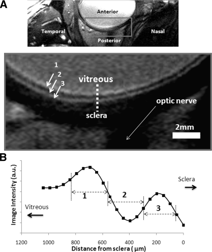
Figure 2.
(A) A bSSFP MRI of the human eye with 100 × 200 × 2000-μm resolution at 3 Tesla. The vitreous appeared relatively bright and the sclera behind the retina appeared relatively dark. Laminar structures within the retina/choroid show three layers of hyper-, hypo, and hyperintense strips (layers 1–3). (B) A spatial profile taken across the retinal thickness from vitreous to sclera.

