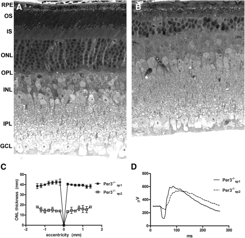
Figure 1.
Representative images of 10-month-old normal (A) and degenerate (B) retinas observed among Per3−/− mice. Note the dramatic thinning of the outer layers of the retina. OS, outer segments; IS, inner segments; ONL, outer nuclear layer; OPL, outer plexiform layer; INL, inner nuclear layer; IPL, inner plexiform layer; GCL, ganglion cell layer. ONL thickness as a function of retinal eccentricity is shown in (C) for 10-month-old animals (n = 3). Remaining animals in the colony were screened by ERG and separated into two subpopulations, which we designated Per3−/−sp1 and Per3−/−sp2 (D). Normal animals (Per3−/−sp1) had a robust a-wave, whereas affected animals (Per3−/−sp2) showed significant a-wave attenuation. ERGs are averages of four to six animals between 6 and 8 weeks of age that were dark adapted for 2 hours and recorded after a single flash of intensity sufficient to evoke the maximum response.

