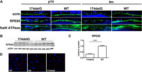
Figure 6.
(A) Cryosections of eyes from wild-type and Mfrp174delG mice were stained with antibodies against ezrin, Na/K ATPase, and RPE65 (green). (arrowheads) Examples of RPE nuclei (blue). Images are oriented such that the apical domain of the RPE is below the RPE nuclei. Na/K ATPase staining basal to RPE nuclei is from choroidal cells. Images are scaled so that staining intensity is roughly equivalent and protein localization can be easily compared, and they are not quantitative. (B, C) Western blot analysis demonstrated significant downregulation of RPE65 in 7-week-old Mfrp174delG mice. This was likely due to random assortment of an RPE65 polymorphism that decreased the stability of the protein. (D) ZO-1 staining of primary RPE explants. ZO-1 (red) was properly localized to the cell periphery in both control and mutant explants.

