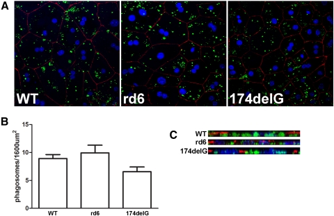
Figure 8.
In vitro phagocytosis assays were conducted to evaluate the overall capacity of mutant RPE cells to internalize OS particles. (A) Representative images showing primary RPE cultures after 2-hour incubation with FITC-labeled purified outer segments (green). Cells are immunolabeled for ZO-1 (red) to indicate cell junctions and stained with TO-PRO3 to label nuclei (blue). (B) Averaged results from four to six fields are shown and are not significantly different (P = 0.079, ANOVA). (C) X-projections were constructed from the confocal stacks to verify that the observed OS particles were internalized and basal to the ZO-1 staining.

