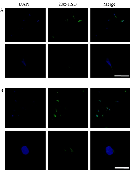
Figure 8.
Bovine 20α-HSD expression in cultured bovine and rat corpus luteum cells by immunofluorescence. Counter staining was performed with DAPI (blue). Bovine 20α-HSD expression was detected by Alexa 488 (green). The merged picture shows blue and green colors. (A) Bovine luteal cells. (B) Rat corpus luteal cells. White bars=100 μm.

