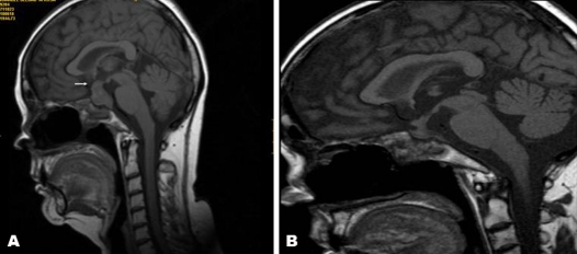
Figure 3.
MRI scan of the pituitary gland (A) before and (B) after corticosteroid treatment. Enlarged pituitary gland with infundibular thickening (arrow) and loss of the hyperintense signal of the posterior pituitary on T1-weighted images (A) that showed a marked reduction of the pituitary size 10 weeks after 3 days of intravenous methylprednisolone bolus (B).

