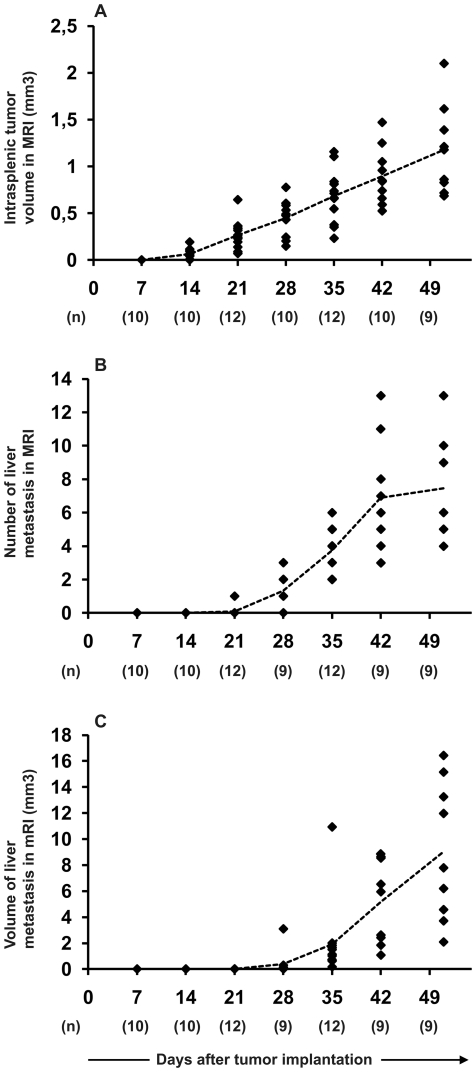
Figure 3. Validation of HT29 cell line in the metastatic colorectal cancer model.
1x10e6 cells were injected into the spleen of 125-day-old SCID mice and intrasplenic tumor growth (A), number of metastatic lesions in the liver (B), and total volume of liver metastases (C) were assessed weekly with MRI. Each dot represents an individual animal and mean of each time point is marked with dotted line. Number of animals analyzed at each time point is presented in parentheses.

