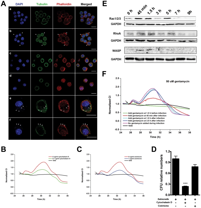
Figure 3. Correlation of Salmonella infection TCRP of intestinal cells and cytoskeleton-associated morphological dynamics.
(A) Immunofluorescence images of HT-29 cells infected with S. typhimurium. (a) uninfected cells; (b) cells at 45 min post-infection. Actin-containing stress fibers are visible, as large accumulations of polymerized actin surrounding invading bacteria (arrows). (c and d) Cells at 1.5 h and 3 h post-infection. Actin cytoskeleton had normal architecture. (e) Cells at 5 h post-infection. Arrows, membrane ruffling and Salmonella-containing vacuoles (SCV). (f) Cells at 7 h post-infection. Arrows, membrane rupture and release of intracellular bacteria. (B) After pretreatment with cytochalasin D for 1.5 h, HT-29 cells were infected with S. typhimurium (MOI = 200). (C) After pretreatment with colchicine for 1.5 h, HT-29 cells were infected with S. typhimurium (MOI = 200). (D) HT-29 cells were pretreated with mock medium, cytochalasin D or colcocine for 1.5 h and infected with S. typhimurium at an MOI of 200. The number of invasive bacteria was determined by gentamycin protection assay. Data are mean ± S.E.M with the number of intracellular bacteria in mock-treated cells set as 1.0. ***, p<0.001 (Student's t-test). (E) Expression levels of cytoskeleton-associated proteins during Salmonella infection measured by western blotting. (F) Profiles of Salmonella infection after treatment of HT-29 cells with gentamycin (50 µg/ml) during infection. Arrows in B, C, F: bacterial addition points. Representative curves are an average of four replicate wells.

