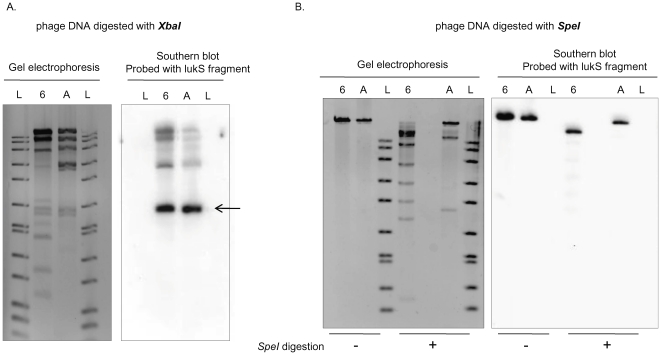
Figure 3. Southern blot analysis of PVL phage genomic DNA from strain 68111 and strain ATCC49775.
Phage DNA from MSSA68111 (Lanes designated “6”) and from strain ATCC49775 (Lanes “A”) was digested with either XbaI (A.) or SpeI (B.). In both A and B, the panels on the left are photographs of ethidium bromide-stained gels; the panels on the right are Southern blots hybridized with the lukS-PV probe. 1 kb DNA ladders (Lanes “L”) are shown. The lukS-PV probe hybridized to identical 2 kb fragments in XbaI -digested phage DNA from both strains (A, arrow) whereas SpeI-digestion produced different sized lukS-PV fragments (B.).

