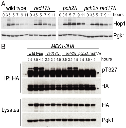
Figure 2. Pch2 and Rad17 promote Hop1 and Mek1 activation.
(A) Western blot analysis of WT, rad17Δ, pch2Δ and pch2Δ rad17Δ at indicated time points after transfer to SPM using α-Hop1 antibody. Pgk1 Western blot was used as the loading control. The phosphorylated isoforms of Hop1 are detectable as slow-moving species. (B) Mek1–3HA immunoprecipitates from WT, rad17Δ, pch2Δ and pch2Δ rad17Δ at indicated time points were analyzed by Western blot using α-phospho-Akt substrate (recognizing pT327 of Mek1) and α-HA antibodies. *IgG heavy chain. Cell lysates were analyzed by Western blot using α-HA and α-Pgk1 antibodies.

