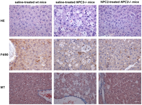
Figure 5. Histochemical and immunohistochemical analysis of NPC2 replacement therapy in murine liver sections.
Staining of representative tissue sections of 87 days old saline treated wild type mice (left panels), saline treated NPC2−/− mice (middle panels), and NPC2 treated NPC2−/− mice (right panels). Hematoxylin-eosin (H&E) staining (first row), immunohistochemical localisation of antigen F4/80 positive macrophages (brownish) (second row), and Masson's trichrome staining to detect collagen (blue) (third row). Lipid laden macrophages (Kupffer cells) are clearly visible and prominent in liver section from saline treated NPC2−/− mice, whereas only a minority of the macrophages in liver sections from NPC2 treated NPC2−/− mice are correspondingly loaded. Data are representative of three separate experiments. n = 3 animals in each experimental group. Scale bars represent 80 µm.

