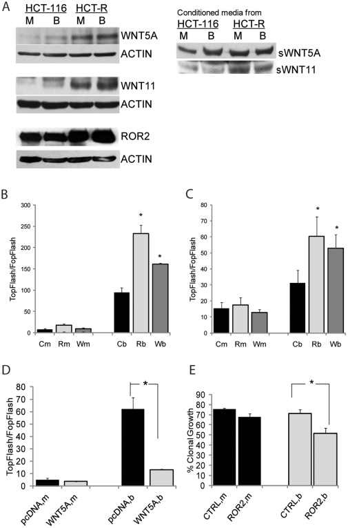
Figure 1. HDACi-resistant HCT-R cells suppress Wnt/beta-catenin activity by overexpressing mediators of noncanonical Wnt signaling.
(A) Representative western blot analyses of CC cells and their conditioned media after exposure to mock treatment (M) or 5 mM butyrate (B) for 19 hrs. (B and C). Silencing of WNT5A and ROR2 expression increases Wnt/catenin transcriptional activity of HCT-116 (B) and HCT-R (C) cells. Cells (50,000/well) were co-transfected with reporters for Wnt/catenin transcriptional activity (TopFlash or FopFlash, 50 ng/well), pRL-TK as a control for transfection efficiency, and control, ROR2, or WNT5A siRNAs (10 pmol/well). Nucleic acids were introduced in the cells via a reverse transfection protocol with Lipofectamine 2000 in 96-well plates. At 31 hrs post-transfection, cells were exposed for 20 hrs to mock (m) or 5 mM butyrate (b) treatment. Wnt transcriptional activity was calculated as the ratio of the activity of TopFlash (with wild type Lef/Tcf binding sites) and FopFlash (with mutant Lef/Tcf binding sites). Each transfection was performed in duplicate wells; data represent the mean from results of three experiments. (D). Overexpression of WNT5A suppresses canonical Wnt transcriptional activity of HCT-116 cells. The co-transfection of HCT-116 cells (50,000/well) with the Wnt activity reporters TopFlash or FopFlash (50 ng per well), and pcDNANeo3.1 or WNT5A-expressing construct (150 ng per well) were carried out through a reverse transfection with Lipofectamine (0.7 µl/well) in 96-well plates. Post-transfection, cells were exposed to mock treatment (m) or 5 mM butyrate (b) for 20 hrs. Each transfection was performed in duplicate wells; data represent the mean from results of at least three experiments. (E) Suppression of ROR2 expression increases the sensitivity of the HDACi-resistant HCT-R cells to the growth-suppressive effect of butyrate. HCT-R cells (106) were nucleofected with 175 pmol of ROR2 (Invitrogen) or control siRNA, and 500,000 cells were plated per well in a 6-well plate. At 24 hrs, cells were exposed to mock treatment (m) or 5 mM butyrate (b) for 17 hrs. At 48 hrs post-nucleofection, cells were plated at 100 or 200 per well in triplicates. Percent clonal growth was calculated as the number of colonies arising from a single cell suspension of 100 cells. The experiment was repeated three times and the results are the mean from the data of the three experiments. Statistically significant differences are noted by stars.

