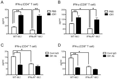
Figure 9. Activation of IFN-γ from lung T cells by lung macrophages during immune responses.
(A, B) Lung CD4+ (A) or CD8+ (B) T cells were isolated from influenza virus challenged WT mice and stimulated with H1N1-pulsed lung-derived macrophages from either WT or IFNαR−/− mice. Cells were co-cultured with plate-coated rDll1 (2.5 µg/ml) or PBS control. (C, D) Lung CD4+ (C) or CD8+ (D) T cells were isolated from influenza virus challenged WT mice and stimulated with H1N1-pulsed lung-derived macrophages from either WT or IFNαR−/− mice. Cells were co-cultured with control IgG or anti-Dll1 Ab (20 µg/ml). Cytokine level of IFN-γ was measured using a Luminex system. Data shown are mean ± SEM and are from a representative experiment of 3 independent experiments. Each time point represents 4 mice per group. *P<0.05, ** P<0.01.

