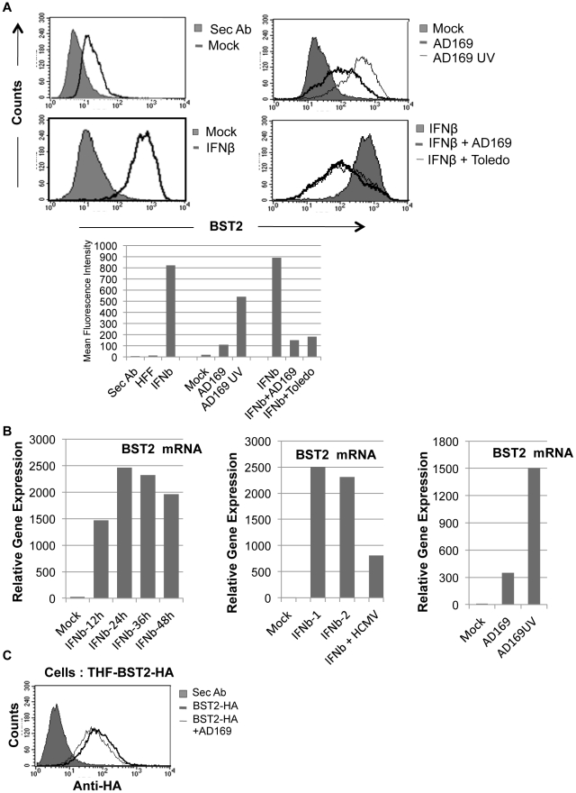
Figure 3. HCMV induces BST2 independent of IFN, but inhibits IFN-dependent induction.
A) Expression of BST2 monitored by flow cytometry using anti-BST2 (HM1.24) antibody is shown in all panels except for the shaded graph in the top left panel that shows staining of secondary antibody alone. Lower panels: Uninfected or HCMV-infected HFFs treated with 500 U of IFNβ for 24 hrs. Right Panels: HFFs infected with indicated viruses (MOI = 3) for 24 hrs. The levels of BST2 are graphically represented below. B) Left panel: qPCR of BST2 mRNA in HFFs treated with 500 U of IFNβ over indicated times. Middle panel: qPCR of BST2 mRNA in HFFs treated with 500 U of IFNβ (two independent experiments) for 24 hours or simultaneously infected with HCMV-AD169 (MOI = 3) for the same amount of time. Right panel: qPCR of BST2 upon infection of HFFs with live or UV-inactivated AD169. C) Flow cytometry of BST2-HA146-expressing THFs uninfected or infected with HCMV-AD169 using anti-HA antibody.

