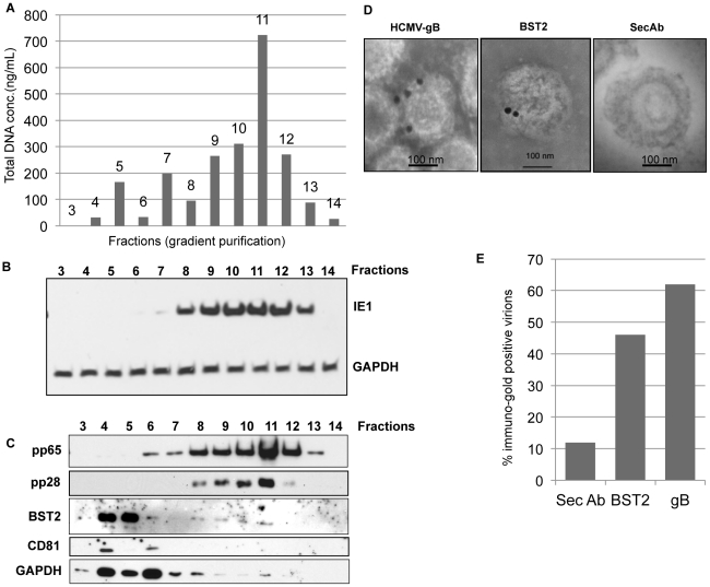
Figure 7. BST2 is present in HCMV virions.
A) HCMV preparations were separated by a discontinuous (5–50%) Nycodenz gradient and equal-sized fractions were isolated prior to spectrophotometric analysis of total DNA. B) Immunoblot for IE1 or GAPDH of HFFs infected with each of the fractions. C) Fractions were probed for the viral proteins gB, pp65 and the cellular proteins BST2 (PNGaseF treated), CD81 and GAPDH by immunoblot. D) Immunoelectron microscopy analysis of purified virions for BST2 and HCMV gB. 20 nm gold conjugated secondary antibody was used to detect BST2 and gB. E) Percentage of immunogold positive virions counted from multiple plates for each treatment.

