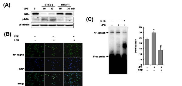
Figure 2.
Effect of BTE on LPS-induced NF-κB signaling in BMM. The cells were pretreated with BTE (100 μg/ml) and then stimulated with LPS (1 μg/ml) for times indicated. (A) IκBα phosphorylation/degradation was evaluated by Western blotting. The LPS-induced IκBα phosphorylation/degradation was dramatically inhibited by coincubation LPS plus BTE. (B) Nuclear translocation of NF-κB/p65 was visualized by immunofluorescence. Coincubation with LPS plus BTE inhibited the nuclear translocation of NF-κB/p65. (C) An EMSA on nuclear extracts of BMM by using a consensus oligonucleotide for NF-κB binding was performed. BTE inhibited LPS-induced NF-κB DNA-binding activity. Bars represent the mean ± SD from three different experiments. (*p < 0.05, compared to unstimulated cells; #p < 0.05, compared to LPS stimulation). BTE, black tea extract; BMM, bone marrow-derived macrophage.

