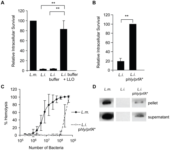
Figure 3. LLO is sufficient to induce the entry of noninvasive L. innocua into HepG2 cells.
(A) HepG2 cells were infected with WT L. monocytogenes (DP10403S, L.m.), L. innocua (L.i.), or L. innocua treated with 1 mM nickel (II) chloride coating buffer in the absence (L.i. buffer) or presence of 5 µM LLO (L.i. buffer + LLO) at MOI = 20 for 30 min at 37°C. Gentamicin was added for 1 h and the intracellular CFUs were enumerated. Results were the mean ± SEM (n≥3) and expressed relative to L. monocytogenes. (B) HepG2 cells were infected with L. innocua (L.i.) or L. innocua phly/prfA* (L.i. phly/prfA*) for 60 min at 37°C (MOI = 100). Gentamicin was added for 30 min and the intracellular CFUs were enumerated. Results were the mean ± SEM (n≥3) and expressed relative to L. i. phly/prfA*. (C) Hemolytic activities of L. monocytogenes (DP10403S) and L. innocua phly/prfA* measured at 37°C for 30 min, pH 7.4. Results were the mean ± SEM (n≥3). (D) Equivalent amounts of L. monocytogenes and L. innocua lysates and proteins precipitated from their culture supernatants were analyzed by western blotting using an anti-LLO primary antibody. A representative experiment (of 3) is presented.

