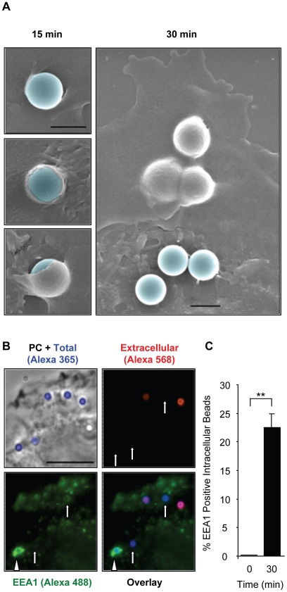
Figure 8. LLO-coated beads are internalized into EEA1 positive endosomes.
(A) HepG2 cells were incubated at 37°C with BSA/LLO-coated beads for 15 or 30 min, washed, fixed, and processed for scanning electron microscopy analysis. Scale bar = 1 µm. (B) HepG2 cells were incubated for 30 min at 37°C with BSA/LLO-coated beads, washed, fixed, and the extracellular beads were labeled with anti-BSA antibodies and a fluorescent secondary antibody (red). After permeabilization, EEA1 was labeled using anti-EEA1 antibodies and a fluorescent secondary antibody (green). Scale bar = 10 µm. PC = phase contrast. Arrows point out internalized beads and the arrowhead an internalized bead that massively recruited EEA1. (C) Results were expressed as the mean ± SEM (n≥3) percentage of intracellular beads that recruited EEA1.

