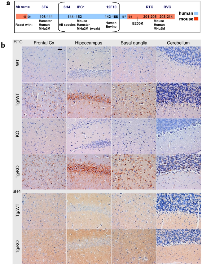
Figure 3. PrP immunoreactivity in the brains of TgMHu2ME199K mice.
a: Epitope mapping of α PrP antibodies used in these experiments. b: Four µm thick sections of formalin fixed, paraffin embedded brains from 8 months old TgMHu2ME199K on wt and ablated background, as compared to wt and PrP ablated mice were tested for disease related PrP immunoreactivity in diverse brain regions with both α PrP pAb RTC and α PrP mAb 6H4. The figure represents at least 3 samples from each group, and depicts intracellular PrP staining with RTC for the sick Tg mice. Scale bar in the upper left panel indicates 20 µm.

