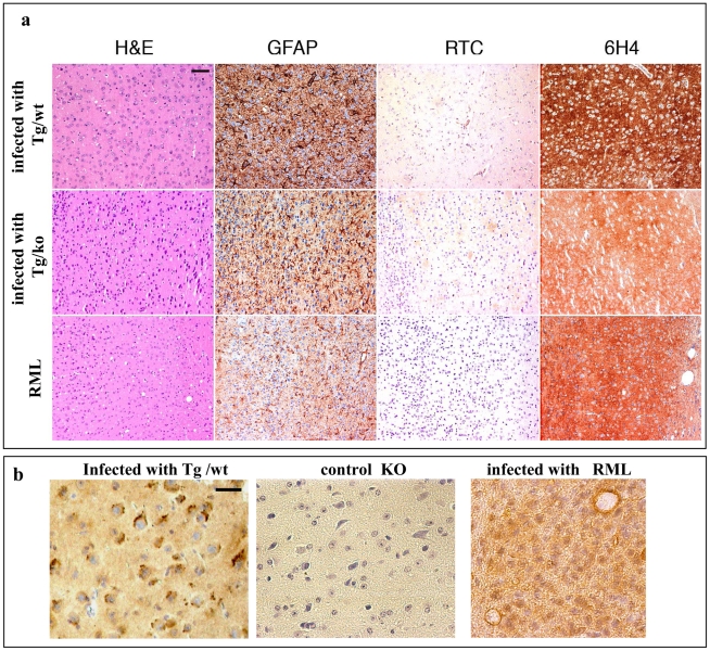
Figure 8. Transmission of TgMHu2ME199K prions to wt mice: pathology.
a: Frontal cortex sections of mice infected with RML or with TgMHu2ME199K prions on a wt or ablated background, analyzed for prion pathological properties, spongiosis, gliosis, and disease related PrP immunoreactivity with pAb RTC and mAb 6H4. Scale bar in upper left image indicates 50 µm. b: RTC immunoreactivity in the absence of formic acid for brains infected with TgMHu2ME199K/wt or RML, as well as for naïve PrP ablated mice. Picture shows different pattern of immunoreactivity for both infected samples. Scale bar in upper left image indicates 20 µm.

