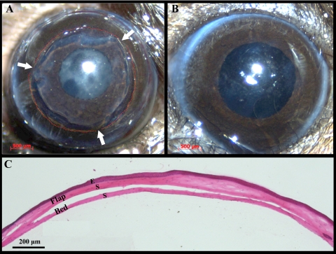
Figure 3.
Lamellar dissection surgery in the murine cornea to transect nerves. (A) Cornea immediately after the lamellar dissection surgery. Red dashed lines: perimeter of the dissection area; arrows: flap hinges. Some retraction of the flap edge is evident. (B) The same cornea 2 weeks after surgery. No signs of scar formation, inflammation, or neovascularization are present. (C) Hematoxylin and eosin staining of a sagittal corneal section immediately after lamellar dissection surgery. Viscoelastic was injected in the flap-bed interface to facilitate flap area measurements. E, epithelium; S, stroma.

