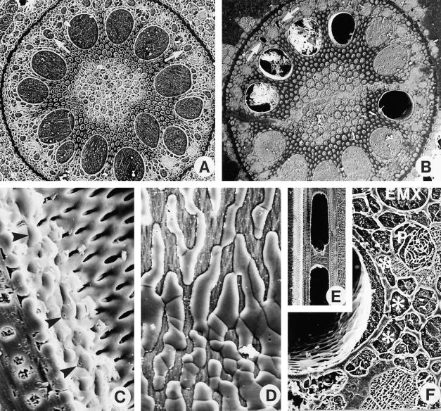
Figure 1.
Cryo-scanning electron microscopy images of field-grown corn roots. The roots were frozen while still attached to the plants, and the frozen pieces were cryoplaned, lightly etched at −90°C, coated with Al, and observed in the secondary electron mode. A, B, and F, Transverse faces. C, D, and E, Longitudinal faces. A, Root frozen at 6 h has no embolized xylem vessels. Magnification is ×112. B, At 8 h, 5 of the 12 LMX vessels were embolized in this root. Some frozen debris from the cryoplaning has fallen into three of the embolized vessels. Magnification is ×104. C, Root frozen at 10 h. The face of the block passes through an embolized LMX vessel that is refilling (as indicated by the convex shape of the frozen drops). Some pits in the lateral wall of this vessel (toward the right of the micrograph) are not yet flooded, but drops of xylem sap were entering the vessel lumen through pits closer to the left of the micrograph (arrowheads). The block face includes a section of part of the longitudinal wall of the vessel showing the bordered pits and the thin primary wall in the floor of each pit. Magnification is ×1600. D, A face view of the lumen side of the lateral wall of an LMX vessel in a root frozen at 14.5 h. Sap droplets have coalesced into rivulets, which obscure the underlying pits. Magnification is ×1680. E, Root frozen at 14.5 h. A column of water in the vessel is sandwiched between two bubbles of gas. Water movement to or from such a water mass must be radial. Magnification is ×112. F, Root frozen at 14 h showing an LMX vessel containing a lens of xylem sap on the side facing the endodermis. Asterisks indicate connecting vessels of a branch root. P, Sieve tube; EMX, early metaxylem vessel. Magnification is ×720.

