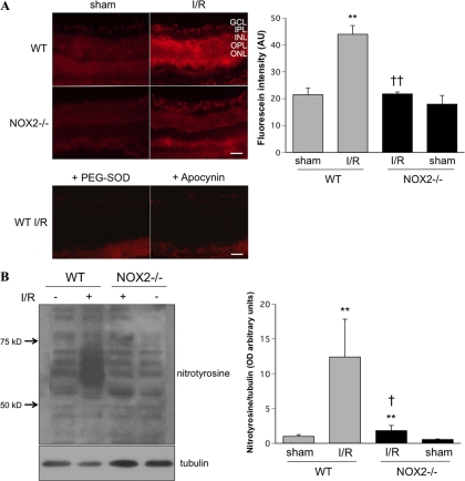
Figure 2.
I/R induced-ROS formation was blocked by NOX2 deletion. (A) DHE imaging of superoxide formation at 6 hours after I/R showed increased DHE reaction in WT I/R mice. This effect was blocked in the NOX2-deficient mice and when sections were pretreated with SOD (400 U/mL) or apocynin (1 mM) (n = 4; **P < 0.01 vs. sham; ††P < 0.01 vs. WT I/R). (B) Western blot analysis shows a marked increase of nitrotyrosine formation in the WT I/R retina compared with WT sham. Knocking out NOX2 blocks this I/R effect (n = 4; **P < 0.01 vs. sham; †P < 0.05 vs. WT I/R). Scale bar, 50 μm. GCL, ganglion cell layer; IPL, inner plexiform layer, INL, inner nuclear layer; OPL, outer plexiform layer; ONL, outer nuclear layer.

