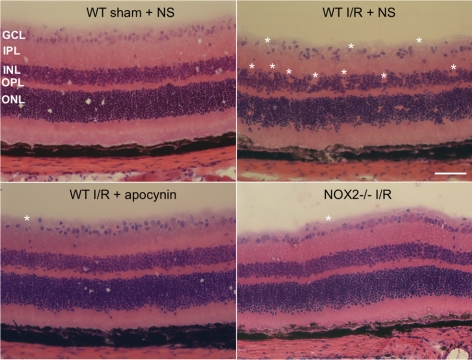
Figure 3.
Apocynin treatment and NOX2 deletion preserve retinal morphology during I/R. Imaging of hematoxylin and eosin–stained retinal sections from I/R mice shows loss of the cells in the GCL and inner nuclear layer (*areas of distortion/missing cells) at 7 days after I/R in WT mice. This effect of I/R is markedly reduced in WT mice treated with apocynin and in NOX2−/− mice. Scale bar, 50 μm. NS, normal saline; GCL, ganglion cell layer; IPL, inner plexiform layer; INL, inner nuclear layer; OPL, outer plexiform layer; ONL, outer nuclear layer.

